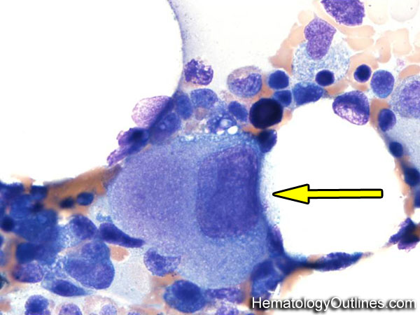
Increased numbers in bone marrow: MDS Reactive increase secondary to peripheral destruction of platelets Post Chemotherapy Toxin-induced
› Classic Immunophenotype:
CD41+
CD42+
CD61+
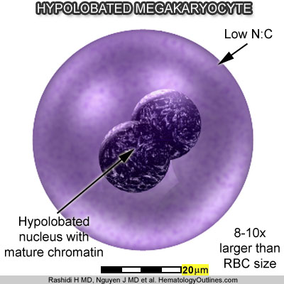
› Cartoon Image:
Click and drag for direct comparison
› Misc:
Hypolobated Megakaryocytes may be dysplastic and associated with a myelodysplastic syndrome (MDS) or newly forming megakaryocytes that are the result of a peripheral destruction of platelets (e.g. ITP)